
Health Answers Made Simple Before Your Diagnostic Test
Explore clear, patient-friendly articles written by our radiology team to help you understand diagnostic imaging, prepare for your appointment, and make confident healthcare decisions. From fetal ultrasound guidance to MRI preparation tips, Sai Dhwani's medical insights are here to support your diagnostic journey.
Helpful Guides for Confident Diagnostic Decisions
Evidence-based articles from Sai Dhwani's radiology team, designed to help you understand diagnostic imaging procedures, prepare for your appointment, and work confidently with your referring physician.
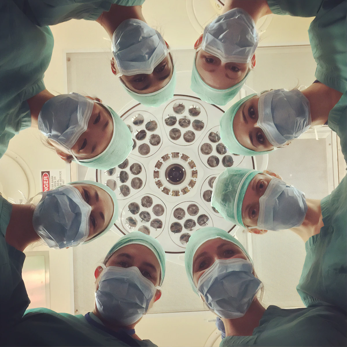
Understanding Fetal Ultrasound: A Complete Guide for Expectant Parents
A comprehensive explanation of fetal ultrasound imaging, why it is important during pregnancy, what to expect during your appointment, and how Dr. Madhura Deshmukh uses advanced ultrasound technology for accurate fetal assessment.
Read Full ArticleThe Importance of Regular Mammography Screening

Learn why annual mammography is essential for early breast cancer detection, what the procedure involves, and how modern mammography at Sai Dhwani supports your women's health.
Read ArticleHow to Prepare for Your Fasting Blood Test
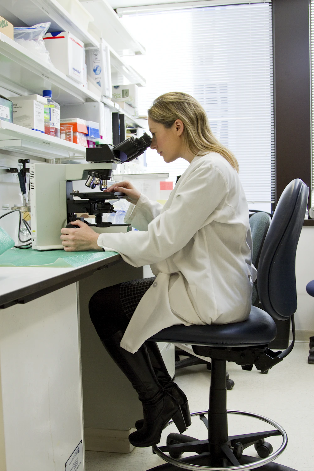
Practical guidance on fasting requirements, what to expect during your blood draw, medication considerations, and how proper preparation ensures accurate test results.
Read ArticlePreparing for Your Abdominal Ultrasound: A Step-by-Step Guide
Understand what to eat or drink before your ultrasound, comfortable clothing tips, what the radiologist will examine, and how to feel confident during your diagnostic appointment.
Read ArticleCT Scan vs MRI: Which Imaging Technology is Right for You?
A detailed comparison explaining how CT and MRI work differently, their benefits, which condition each imaging type typically diagnoses, and what to expect during each procedure.
Read ArticleUnderstanding Your Radiology Report: A Patient's Guide
Demystify medical terminology in your radiology report. Learn how to read your imaging results, what findings mean, and how to discuss your report with your doctor for informed next steps.
Read ArticlePatient Stories

Voices of Trust
Real stories from real patients. Read how Dr. Madhura Deshmukh’s accurate diagnosis, advanced imaging expertise, and compassionate approach have helped patients get clarity, confidence, and the right direction for their treatment.
4.9
120+ Reviews
A
ARVIND SAINI
Sai Dhwani is the best diagnostic centre I have ever seen.Good & very co-operative staff. The place is very clean. I went for my sonography and my experience is really good.Dr...Read More
Y
The Yogic Bliss
Really loved the service at Sai Dhwani diagnostics center which is in Koper Khairane sector 9 just opposite to Balaji theatre. Dr. Madhura Deshmukh is so kind and so good...Read More
H
Himani Saxena
Had a great experience here. Dr. Madhura Deshmukh is kind and professional. The center is clean, hygienic, and equipped with modern instruments. Highly recommended!
S
SNEHA GAJBHIV
Really Good service At Sai dhwani centre.Dr.Madhura Deshmukh ma'am is very polite & Knowledgeable Person makes patient comfortable.staff is Good &...Read More
V
Vineeta khatri
Very polite doctor and staff. Clean and well maintained place. Will surely recommend.
Read All Reviews on Google
Why You Can Trust Sai Dhwani's Radiology Insights
Our blog content is written by Dr. Madhura Deshmukh and her team of experienced radiologists. Every article is grounded in medical evidence, designed to empower patients with accurate information, and aligned with the highest standards of patient education.
Written by Radiologists
All blog content is created by Dr. Madhura Deshmukh (MBBS, DMRE—10+ years experience) and her clinical team. You're reading insights from specialists who diagnose and interpret imaging every day.
Accurate Medical Information
Our articles are based on current medical evidence, diagnostic imaging best practices, and guidelines from leading radiology associations. We prioritize accuracy and clarity over sales messaging.
Built to Help You
Every article is designed to help patients understand their diagnostic journey. We explain medical concepts in simple language and empower you to ask informed questions of your physician.
Trusted by 263+ Patients
Sai Dhwani is rated 4.9/5 by verified patients. Our commitment to clear communication, accurate diagnosis, and patient education has earned the trust of individuals and referring doctors across Navi Mumbai.
Frequently Asked Questions
Answers to Common Patient Queries About Colorectal, Breast & General Surgeries in Thane
Which is the best radiology and diagnostic center in Kopar Khairane, Navi Mumbai?
Sai Dhwani Lab Center, located at Shop No. 5, Ground Floor, Balaji Exotica, opposite Balaji Movieplex, Sector 9, Kopar Khairane, is one of the most trusted radiology and diagnostic centers in Navi Mumbai. Led by Dr. Madhura Deshmukh (MBBS, DMRE) with over 10 years of specialized experience, Sai Dhwani offers comprehensive diagnostic services including ultrasound, fetal imaging, Doppler studies, echocardiography, and lab tests. Rated 4.9 stars by 263 verified patients, our center is recognized for accurate reporting, advanced imaging technology, and compassionate patient care across Kopar Khairane, Ghansoli, and Navi Mumbai.
What is a fetal anomaly scan, and when should I get it done during pregnancy?
A fetal anomaly scan, also called a TIFFA scan (Targeted Imaging for Fetal Anomalies), is a detailed ultrasound examination performed between 18 to 22 weeks of pregnancy. It carefully examines the baby’s brain, spine, heart, kidneys, limbs, and other internal organs to detect any structural abnormalities or developmental concerns early. At Sai Dhwani Lab Center, Dr. Madhura Deshmukh performs fetal anomaly scans in 2D, 3D, and 4D formats with expert interpretation and clear parental guidance. Early detection through a TIFFA scan helps doctors and parents plan appropriate care and make informed decisions throughout the pregnancy journey.
What is the difference between a 2D, 3D, and 4D ultrasound during pregnancy?
A 2D ultrasound is the standard pregnancy scan that produces flat, two-dimensional black-and-white images used for clinical measurements, fetal development assessment, and medical diagnosis. A 3D ultrasound creates a three-dimensional still image of the baby, showing facial features and body structure in greater detail. A 4D ultrasound is a live 3D video, allowing you to see the baby’s real-time movements, expressions, and gestures. At Sai Dhwani Lab Center, we offer all three formats — 2D, 3D, and 4D — for routine obstetric scans, anomaly scans, fetal echo, and NT-NB assessments. Dr. Madhura Deshmukh will recommend the most appropriate imaging format based on your clinical need and stage of pregnancy.
When should I get a fetal echo done, and what does it check?
A fetal echocardiography (fetal echo) is a specialized ultrasound that examines the structure and function of the baby's heart before birth. It is typically recommended between 20 to 24 weeks of pregnancy. Your doctor may refer you for a fetal echo if there is a family history of congenital heart disease, if a previous scan showed a suspected cardiac abnormality, if you have diabetes or other medical conditions, or as part of a comprehensive fetal assessment. At Sai Dhwani Lab Center, Dr. Madhura Deshmukh performs fetal echo in 2D, 3D, and 4D formats with detailed cardiac assessment and clear reporting to guide your treating doctor and ensure the best care for your baby.
What is a colour Doppler test, and when do doctors recommend it?
A colour Doppler ultrasound is a specialized imaging technique that measures and visualizes blood flow through arteries, veins, and organs in real time using colour-coded imaging. Doctors recommend colour Doppler for a wide range of conditions including monitoring blood flow between mother and baby during pregnancy (obstetric Doppler), detecting blood clots or vascular blockages, assessing kidney blood flow, evaluating liver and portal vein circulation, screening for deep vein thrombosis, and checking carotid artery health. At Sai Dhwani Lab Center, we offer colour Doppler studies of all kinds, including obstetric Doppler, renal Doppler, hepatic Doppler, peripheral vascular Doppler, and more, with expert interpretation by Dr. Madhura Deshmukh.
What is a whole abdomen sonography, and what does it detect?
A whole abdomen sonography is a comprehensive ultrasound examination of all organs within the abdominal and pelvic cavity, performed in a single session. It evaluates the liver, gallbladder, bile ducts, pancreas, spleen, kidneys, urinary bladder, and surrounding structures for any abnormalities. Doctors recommend whole abdomen sonography for unexplained abdominal pain, bloating, nausea, suspected kidney stones, liver disease, gallstones, pancreatic conditions, and routine health screenings. At Sai Dhwani Lab Center in Kopar Khairane, Dr. Madhura Deshmukh performs whole abdomen sonography with detailed reporting and clear findings to support your doctor’s diagnosis and treatment decisions.
Have Questions About Your Diagnostic Test?
Unsure which imaging test you need? Want to understand your test better? Our team is here to guide you. Fill the form to get personalized recommendations and schedule your diagnostic appointment at Sai Dhwani.
Get expert guidance from our radiology team. Fill out the form below to share your health concerns, and we will help recommend the right diagnostic service.