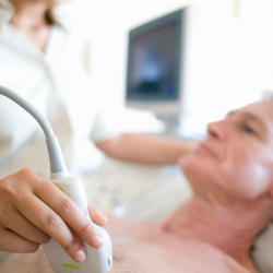
2D Echo
A detailed ultrasound imaging technique for heart evaluation, ensuring accurate cardiac assessments.
2D Echo
2D Echocardiography (2D Echo) is a non-invasive ultrasound imaging technique used to assess the heart’s structure and function in real time. It provides moving images, allowing doctors to evaluate the heart’s size, shape, and pumping efficiency.
Who Needs It?
- Individuals with chest pain, breathlessness, or palpitations
- Patients with high blood pressure or heart disease
- People at risk of stroke or heart failure
- Those undergoing heart surgery or post-treatment follow-ups
Why is it Important?
- Detects heart valve issues & abnormalities
- Helps in early diagnosis of congenital heart defects
- Assesses blood flow and heart function
- Monitors the effectiveness of treatment.

Uses of 2D Echo
Heart Function Assessment
Checks the heart’s pumping ability, detects heart muscle weakness, and evaluates blood flow efficiency.
Valve & Chamber Evaluation
Detects valve disorders, measures heart chamber sizes, and assesses overall cardiac function.
Congenital Heart Defects
Identifies birth-related heart abnormalities and structural defects in infants and adults.
Preparation for 2D Echo
Frequently Asked Questions (FAQs)
Is 2D Echo painful?
No, 2D Echo is a painless procedure. It uses sound waves to create images and does not involve any needles or radiation.
How long does the test take?
The test usually takes 20 to 30 minutes to complete.
Can I drive after a 2D Echo test?
Yes, you can resume normal activities immediately after the test.
Is 2D Echo safe for pregnant women?
Yes, 2D Echo is completely safe for pregnant women as it does not use radiation.
